
Functional anatomy of headache: circle of Willis aneurysms, third cranial nerve and pain
Views: 2381DOI:
https://doi.org/10.48208/HeadacheMed.2011.18Keywords:
Aneurysm, Headache, Oculomotor nerve, Pain, Posterior communicating antery, Internal carotid artery, AnatomyAbstract
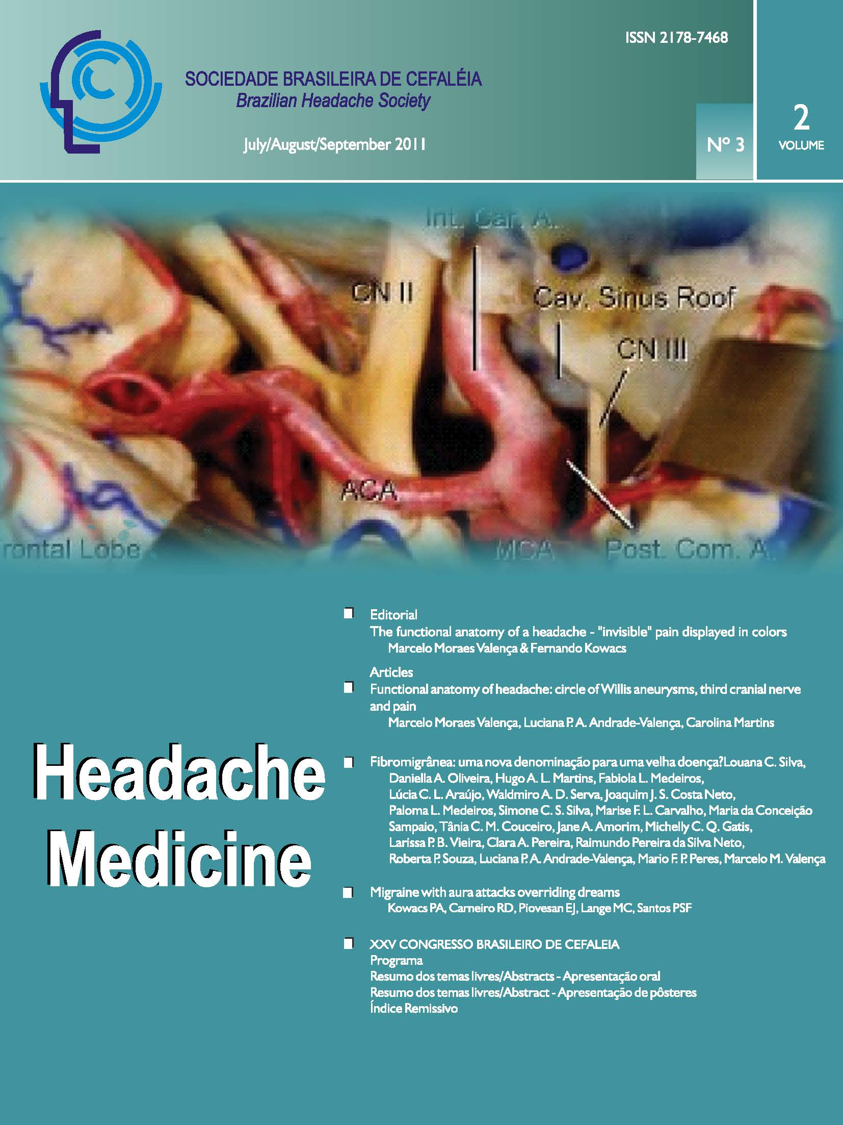
Patients with intracranial aneurysm located at the internal carotid artery-posterior communicating artery (ICA-PComA) often present pain on the orbit or fronto-temporal region ipsilateral to the aneurysm, as a warning sign a few days before rupture. Given the close proximity between ICA-PComA aneurysm and the oculomotor nerve, palsy of this cranial nerve may occurduring aneurysmal expansion (or rupture), resulting in progressive eyelid ptosis, dilatation of the pupil and double vision. In addition, aneurysm expansion may cause compression not only of the oculomotor nerve, but of other skull base pain-sensitive structures (e.g. dura-mater and vessels), and pain ipsilateral to the aneurysm formation is predictable. We reviewed the functional anatomy of circle of Willis, oculomotor nerve and its topographical relationships in order to better understand the pathophysiology linked to pain and third-nerve palsy caused by an expanding ICAPComA aneurysm. Silicone-injected, formalin fixed cadaveric heads were dissected to present the microsurgical anatomy of the oculomotor nerve and its topographical relationships. In addition, the relationship between the right ICA-PComA aneurysm and the right third-nerve is also shown using intraoperative images, obtained during surgical microdissection and clipping of an unruptured aneurysm. We also discuss about when and how to investigate patients with headache associated with an isolated third-nerve palsy.
Downloads
Published
How to Cite
Issue
Section
License
Copyright (c) 2021 Marcelo Moraes Valença, Luciana P. A. Andrade-Valença, Carolina Martins

This work is licensed under a Creative Commons Attribution 4.0 International License.