Functional anatomy of headache: hypothalamus
Views: 2338DOI:
https://doi.org/10.48208/HeadacheMed.2011.24Keywords:
Anatomy, Hypothalamus, Cluster headache, Migraine, DBS, MRIAbstract
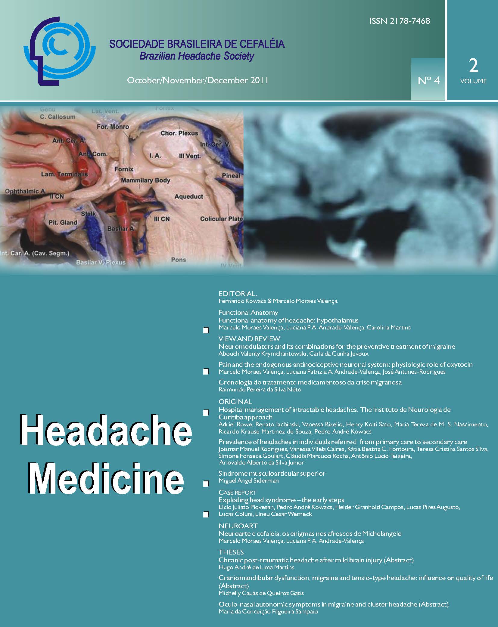
There is now compelling evidence that the hypothalamus exerts a major role in the mechanism of headache triggering. Pain and concomitant changes in the hormonal secretory pattern occur during an attack of headache when hypothalamic structures are involved. During spontaneous migraine or cluster headache attacks activation of the hypothalamus is shown by positron emission tomography. Over the past 10 years a number of patients with refractory chronic cluster headache have received neurostimulation of the posteroinferior hypothalamus as a form of treatment. The clinical use of deep brain stimulation (DBS) is based on the theory of posterior hypothalamic nucleus dysfunction as the cause of cluster headache attacks. In this article the authors review the functional anatomy of the hypothalamic region and its neighborhood, using silicone-injected cadaveric head and MRI. In conclusion, a better understanding of the functional anatomy of the hypothalamus and its neighborhood is imperative for understanding the pathophysiology of several of the primary headaches, particularly migraine and the trigemino-autonomic headaches. Direct stimulation of the posterior hypothalamic region using DBS devices is now the "state of the art" form of treatment indicated for refractory chronic cluster headache. The exact mechanism and the actual region where the DBS may act are still unknown, and studies on the functional anatomy of the hypothalamus are crucial to the progress in this marvelous field of functional neurosurgery.
Downloads
Published
How to Cite
Issue
Section
License
Copyright (c) 2011 Headache Medicine

This work is licensed under a Creative Commons Attribution 4.0 International License.


