
Histomorphometric analysis of mast cells in different regions of human intracranial dura mater
DOI:
https://doi.org/10.48208/HeadacheMed.2019.16Keywords:
Mast cell, Dura mater, Human, Meningeal artery, MigraineAbstract
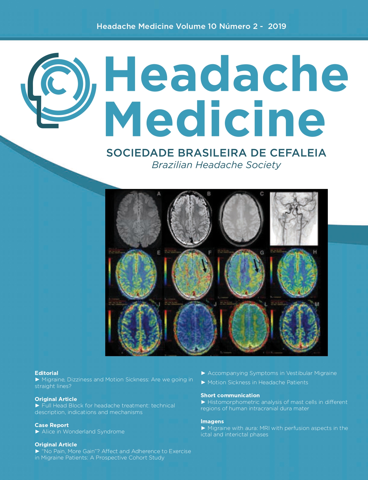
Objective: To analyze mast cell histomorphometry in three different regions of the human intracranial dura mater. Method: Three specimens of dura mater were collected after approval by the Ethics Committee (CAAE No. 57692216.5.0000.5208). Each dura mater was obtained from human cadavers between 7 and 24 hours after death. After collection, the samples were fixed, cut into two fragments and longitudinally placed in the following way: external (periosteum) and internal (meningeal) sides. The fragments (1.5 cm2) were taken from three different regions: proximity of the right middle meningeal artery, the proximity of the left middle meningeal artery and superior sagittal sinus. These fragments were submitted to microtomy (10 μm), stained with 0.1% toluidine
blue and analyzed by optical microscopy. The histomorphometric parameters adopted were: the distance from the mast cells to the vessels, the number and if the mast cells were degranulated. Five fields from each case were analyzed. For this analysis, the Image J 1.52a 2019 software was used. Results: A higher number of mast cells was observed in the periosteal layer when compared with
the meningeal layer (p=0.026). When the distribution of the mast cells was evaluated, we observed that the cells were localized in the proximity of the middle meningeal artery (p<0.05). Conclusion: In human dura mater, the mast cells are localized in the proximity of dural arteries.
Downloads
Published
Issue
Section
License
Copyright (c) 2019 Headache Medicine

This work is licensed under a Creative Commons Attribution 4.0 International License.